Angela Chappell
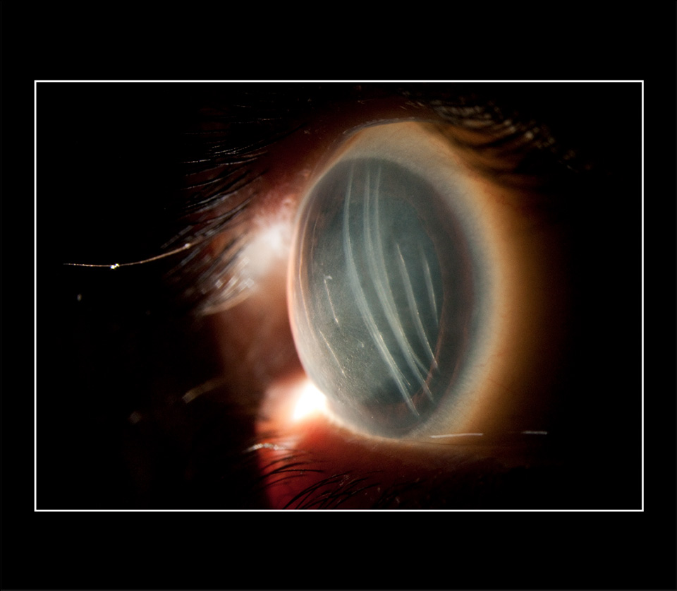
Wolverine Eye/Descemet’s Membrane Folds, 2017
Ophthalmic sclerotic scatter slit lamp photograph
Haag-Streit PBQ900 photo slit lamp
Ophthalmology Department, Flinders Medical Centre
Bedford Park, South Australia, Australia
This external eye photograph was captured using sclerotic scatter lighting. A controlled, decentered slit beam is directed to the corneoscleral junction or limbus, scattering light through the cornea or front window structure of the eye. The linear corneal abnormalities revealed are folds in a deep layer of the cornea which is called Descemet’s membrane. These can be caused by birth trauma, including forceps injury of the eye, or other eye trauma later in life. In this case the clarity of the cornea was adversely affected such that the patient was later given a Descemet’s stripping endothelial keratoplasty or DSEK corneal transplant to restore clear vision.
