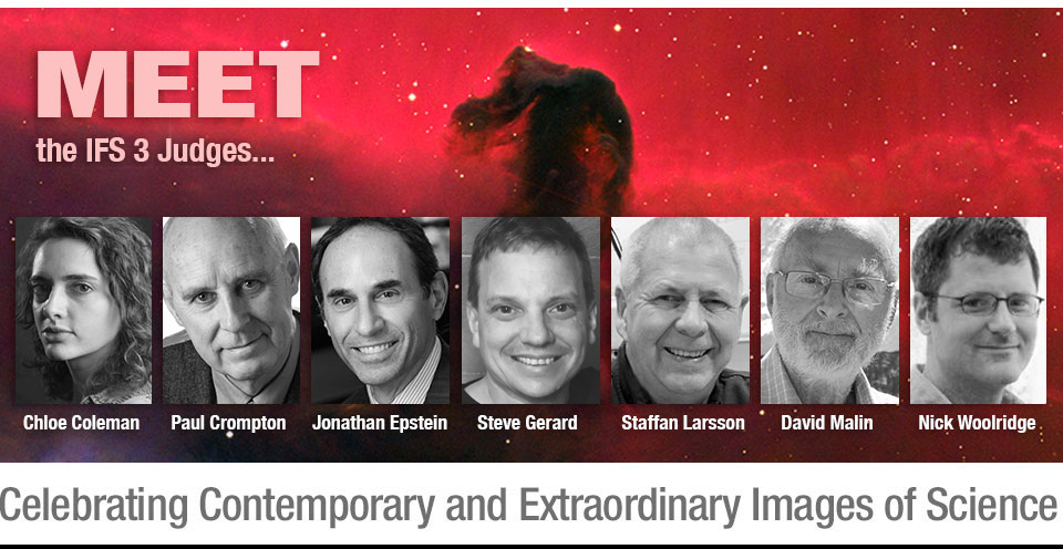
Chloe Coleman
Photo Editor
Washington Post
Washington, DC USA
Chloe Colemanis an award-winning photo editor at The Washington Post, currently working on the international news desk. She is a contributing writer and editor for the paper’s In Sight photo blog, where she has written about and featured contemporary photography, photo books, and exhibitions. Her career in photo editing began as an intern at National Public Radio, followed by her first staff position as a digital photo editor at The Denver Post. She attended the Columbus College of Art & Design and is a graduate of the photojournalism program at the Rochester Institute of Technology. Coleman also serves as a faculty member at The Kalish Visual Editing Workshop and has participated as a juror for several photography contests, including The Magenta Foundation’s Flash Forward competition (2018) and New York Press Photographers Association’s Year in Pictures..
Paul Crompton
Medical Photographer (retired)
University Hospital of Cardiff
Cardiff Wales UK
Paul Crompton studied at Blackpool College of Art and then worked as a trainee medical photographer at the University Hospital of Wales. He quickly progressed to senior photographer, where he earned additional certificates. Crompton has explored different genres of photography and has exhibited his work in Wales’ prestigious Ffotogallery. Crompton became a lecturer at the Cheshire School of Art and Design in 1985 and shortly thereafter became the leader of the vocational photography course. He earned an MA from Derby University in photographic studies, and he was a visiting lecturer in photography at Blackpool College.
Crompton returned to University Hospital of Wales in 1994 as Head of Photography, where he led the conversion from film to digital. He optimized the digital photography services, expanded into ophthalmic imaging, and created initiatives for developing the role of photography within the patient care pathways of dermatology and wound healing. He has written and lectured in the United Kingdom and the United States, and has won a number of awards. He served as a judge for the United Kingdom’s Institute of Medical Illustrators’ awards and for the BioCommunications Association’s exhibitions. In 2014, Crompton was selected as a Louis Schmidt Laureate, a lifetime achievement award from the BioCommunications Association. In 2015 he was awarded an Honorary Fellowship of the Institute of Medical Illustrators. Crompton continues to produce new work and is part of a team documenting outreach projects in Africa. Currently he is working with the charity Mothers of Africa, and Cardiff University’s Phoenix Project, an educational collaboration with the University of Namibia in southern Africa, where he produces multimedia digital stories.
Jonathan Epstein, M.D.
Professor of Pathology, Urology, and Oncology
Reinhard Chair of Urological Pathology
Director of Surgical Pathology
The Johns Hopkins Medical Institutions
Baltimore, Maryland
Dr. Epstein received training in Pathology at Johns Hopkins with a fellowship in Memorial Sloan Kettering Cancer Center. He is the past President of the International Society of Urological Pathology. Dr. Epstein has 898 publications (H-factor – 141), 68 book chapters, and 15 books. He has one of the largest surgical pathology consulting services in the world with approximately 12,000 cases per year, covering the full range of urologic pathology. Dr. Epstein trains 4 genitourinary pathology fellows each year, with 61 fellows trained to date. He has lectured 363 times outside of his institution including 41 different countries. His passion outside of his work is photography in the many countries he has been fortunate to visit.
Steve Gerard
Director
Science Source Images
New York, New York
Steve Gerard has worked at Science Source for nearly three decades as Director and Editor of the science and medical collections. Science Source is a stock photo agency based in New York City that specializes in scientific, medical and high-tech photography. In this role he is responsible for talent acquisition, photographer relations and editing/maintaining the collection of over 500,000 images. Steve also has extensive knowledge of publishing, commercial, television, and film rights-management. At Science Source he has overseen the conversion of the original Science Division (started at Photo Researchers, Inc.) from slides and prints to a fully digital archive. In recent years Steve has worked to acquire and represent content for Science Source’s new video division. Steve has a Bachelors of Science in Biology from SUNY Binghamton.
Staffan Larsson
Imaging Consultant
School Engineering Sciences in Chemistry,
Biotechnology and Health - CBH,
The Jonasson Centre for Medical Imaging/3D Visualization Lab,
KTH Royal Institute of Technology
Stockholm Sweden
Staffan Larsson lives in Stockholm, Sweden and earned an AS degree in photography in 1972 and earned a national certificate in digital image workflow delivery systems. In 2018, he earned his National Swedish Masters Diploma in Photography.
Larsson has over forty-five years of experience in medical and scientific photography. He has worked at Karolinska Institute Medical University and Stockholm County Council Healthcare. In 2009, he joined the Royal Institute of Technology’s School of Technology and Health faculty and Clinical Science Intervention and Technology (CLINTEC) at Karolinska Institute. Larsson has also worked in the arts, photojournalism and in nature. As a photographer his work has illustrated countless science and medicine articles featuring images created using new and interesting ways. He has led workshops and courses for students all across Sweden and the U.S. Larsson was pivotal in the formation of the Lennart Nilsson Award in 1997 and developed initiatives supporting its mission for more than fifteen years. He was an author in The Focal Encyclopedia of Photography fourth edition, wrote a chapter for Laboratory Imaging and Photography in 2016. In 2017, he retired as a research engineer in medical and scientific photography at School of Technology and Health, Royal Institute of Technology where he taught and assisted scientist-working groups as a consultant for communications and public relations. He is currently a consultant to the Image Center located at The Jonasson Centre for Medical Imaging/3D Visualization Lab, KTH Royal Institute of Technology, Stockholm, Sweden
David Malin
Anglo-Australian Telescope and RMIT
Australia
Australian-based photographer and astronomer David Malin was born in England, studied chemistry, and explored photography early on. After working for a multinational company as a chemist using photomicrography, Malin went from exploring the infinitely small to the infinitely far away when he joined the Anglo-Australian Observatory as its photographic scientist in 1975. He remained there for 26 years.
In his photographic laboratory in Sydney, Malin invented new ways of extracting information from astronomical photographs, which lead to the discovery of two new types of galaxies. His novel image enhancement techniques were incorporated into a method of making unique three-color photographs of previously unseen deep space objects.
Malin's photographs have been widely published in books and magazines such as LIFE and National Geographic, and after being recognized for their scientific value, his chromogenic and platinum/palladium prints of the universe have been exhibited in major museums and galleries. His work is part of the collections of museums, institutions and private collectors on an international level.
A well-known lecturer, David Malin has published numerous scientific papers and popular articles on astronomy and photography, as well as nine books, including The Invisible Universe (Bulfinch Press/Little, Brown and Company, 1999), a large format celebration of the beauty of the night sky, and Ancient Light (Phaidon, 2009), a portrait of the universe in black and white.
Nick Woolridge
Director and Associate Professor at Biomedical Communications
University of Toronto
Toronto, Canada
Nick Woolridge received his MSc from the Institute of Medical Science at the University of Toronto in 1996. The topic of his thesis was the development and formative evaluation of a semi-immersive clinical simulation for medical students, which had been funded by SPAR Aerospace. He is an Associate Professor in and Director of Biomedical Communications in the Biology Department of the University of Toronto Mississauga. He conducts research in the development of digital media as instruments of biomedical research, teaching, and patient assistance. He is the co-author of Anatomy 300/303 Interactive Lab Companion, as well as co-author (with Jason Sharpe and Charles Lumsden) of the recent In Silico: 3D Animation and Simulation of Cell Biology with Maya and MEL.