
Bernardo Cesare
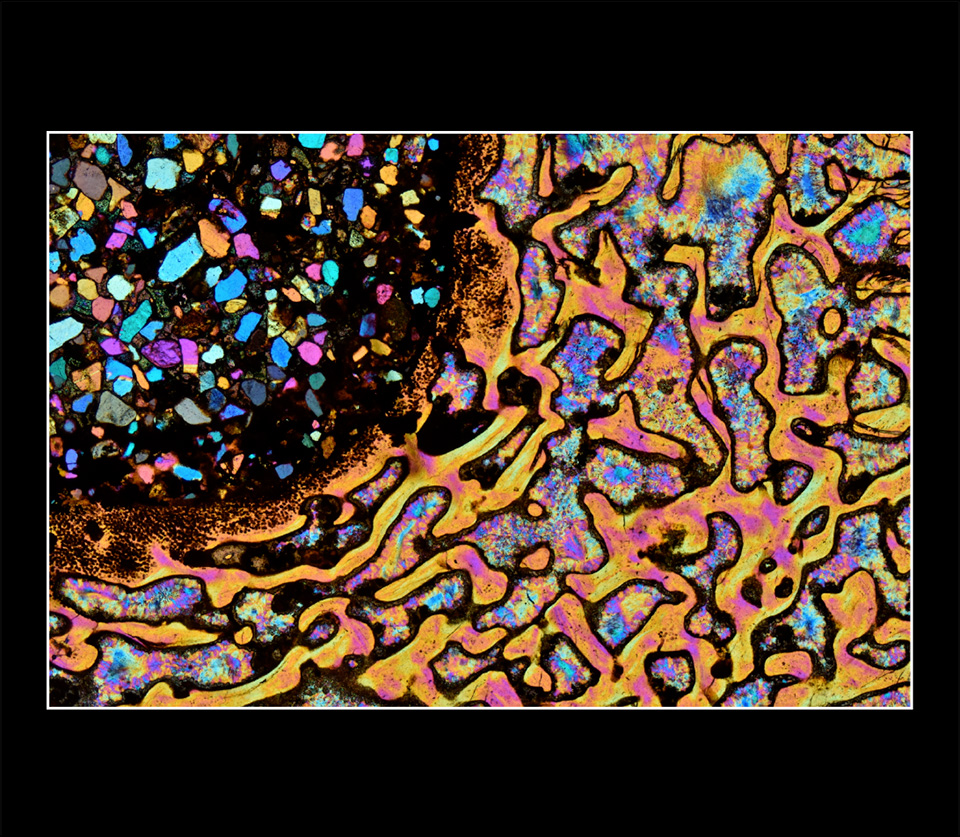
Dinosaur Bone, 2017
Polarized light photomicrograph
Zeiss Axioscope 40 POL polarized light microscope; 2.5x POL objective was used;
polarizers at 90 degrees with a red tint plate in the optical pathway; Nikon D5500 DSLR camera
Geosciences Department, University of Padua
Padua, Italy
This photomicrograph features a 30-micron slice of a dinosaur bone collected in Utah, United States. The image shows the highly porous bone structure; the pores are filled with late chalcedony. The fossil reveals remnants of the bone tissue, and the black dots reveal the shape of former single bone cells. The larger void on the top left was filled with a silt-rich sediment composed of quartz angular clasts. The section of bone visible in the microscope was 5.3 millimeters.