
Alan Prescott
Mitochondrial Turnover in the Developing Mouse Eye, 2018
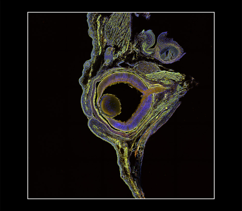
Laser scanning confocal photomicrograph
Dundee Imaging Facility, University of Dundee
Dundee, Scotland, United Kingdom
The tiled images in this photograph were taken from a frozen section of a developing eye harvested from the mitoQC mouse, a transgenic mouse with a pH-sensitive fluorescent mitochondrial signal. Under stable conditions, mitochondria fluoresce both green and red. However, when the process of turnover (self-destruction of damaged mitochondria) begins, the mitochondria are delivered to lysosomes, where the acidic environment of the lysosomes quenches the green fluorescence, guaranteeing specificity of the red autophagy (destruct) signal. Large red dots in the image are mitochondria in lysosomes demonstrating turnover of damaged or worn out mitochondria in active tissues in the developing eye. DNA-labelled nuclei are blue. This mouse model has revealed the distribution of the turnover process in diverse, metabolically active tissues, such as tissue of the kidney, heart, and retina. In addition, this mouse model unveils the tissue architecture as delineated by the distribution of mitochondria. At this developmental stage, the eyelids are closed and the posterior chamber still contains blood vessels to support the developing lens and retina.