
Giulia Bolasco
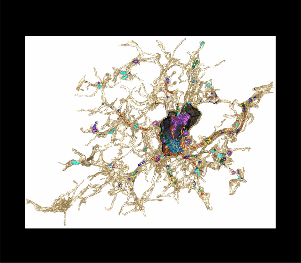
3D Nanostructure of a Microglia Cell Body, Processes, and Internal Organelles, 2018
Confocal photomicrograph
4Serial block-face scanning electron microscope workflow; correlative light and electron microscopy; field of view 32x24x23 microns; anisotropic resolution 5 nanometers in xy orientations and 25 nanometers thick
Microscopy Facility, Epigenetics and Neurobiology
European Molecular Biology Lab
Rome, Italy
This photomicrograph features a five-day-old Arabidopsis thaliana seedling after exposure to latrunculin B, a metabolite isolated from red sea sponge. Latrunculin B blocks the assembly of actin monomers into filamentous structures. The green, red, and blue colors mark distinct regions of the plant cell. The green color marks the filamentous actin (F-actin) cytoskeleton, and the red and blue colors show autofluorescence from chloroplasts and cell walls, respectively. Note that thick actin bundles accumulate in distinct regions within the cytoplasm. The width of the elongated cells is approximately 40 micrometers.