
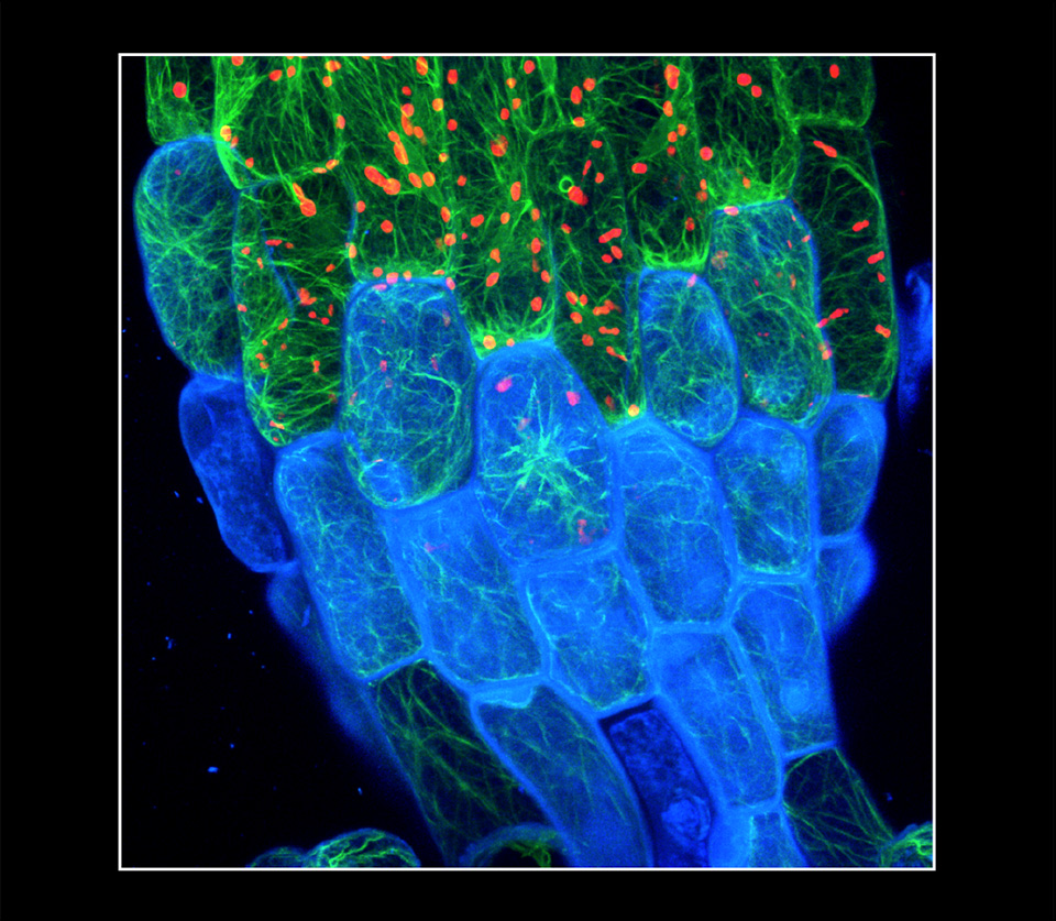
Elison Blancaflor
Chemical Disruption of Actin in Plant Cells, 2018
Confocal photomicrograph
40x water immersion objective; capture magnification ~x400
Noble Research Institute, LLC
Ardmore, Oklahoma, United States
This photomicrograph features a five-day-old Arabidopsis thaliana seedling after exposure to latrunculin B, a metabolite isolated from red sea sponge. Latrunculin B blocks the assembly of actin monomers into filamentous structures. The green, red, and blue colors mark distinct regions of the plant cell. The green color marks the filamentous actin (F-actin) cytoskeleton, and the red and blue colors show autofluorescence from chloroplasts and cell walls, respectively. Note that thick actin bundles accumulate in distinct regions within the cytoplasm. The width of the elongated cells is approximately 40 micrometers.