
James E. Hayden
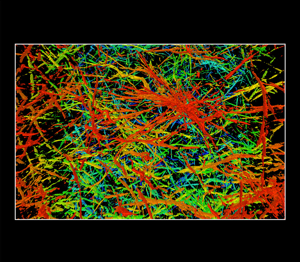
3D Collagen Matrix in Vitro, 2016
Photomicrograph
Two-photon microscope; a composite of 200 stacked images or “slices” through material that is 200 microns thick
The Wistar Institute
Philadelphia, Pennsylvania, United States
A 3D collagen matrix is used in cancer research to support the growth of cell cultures for a more realistic representation of naturally growing tumors. The concentration of collagen is an important part of the extracellular matrix surrounding cancer cells and is partly responsible for the ease with which these cells can invade surrounding tissues. The collagen was visualized with second harmonic generation, a method that allows certain molecules, like collagen, to be seen without direct staining. A depth-coded color lookup table was added to the resulting black and white image to display thickness. Red is closest to the top of the tissue and blue is farthest away.