
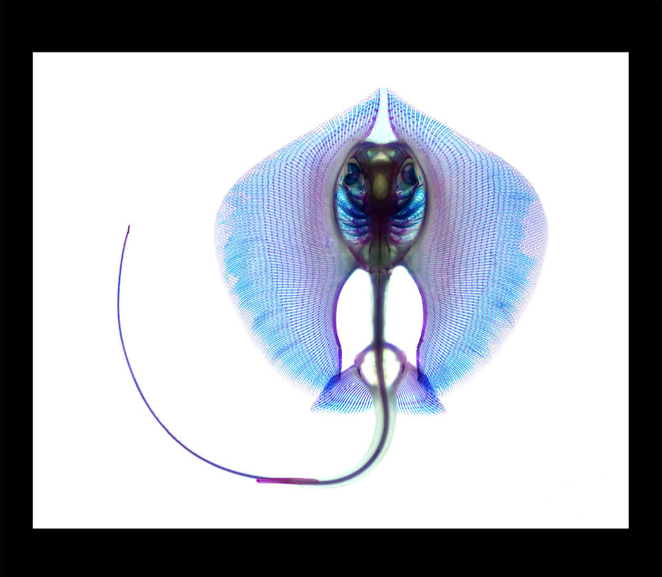
Michelle C. Gilbert
Dasyatis sabina, Atlantic Stingray, 2018
Computational scientific photograph
Canon EOS Rebel T3 DSLR camera; Canon EF-S 18–55mm lens f/3.5–5.6 macro lens;
a composite of numerous images that were focus stacked and stitched together;
images were taken from five overlapping sections of various heights
University of Massachusetts Amherst
Amherst, Massachusetts, United States
This image shows a cleared and stained Atlantic stingray. This specimen was stained using traditional processes. The staining shows the calcified structures as purple and the cartilage as blue. The staining technique enzymatically removes cells to leave behind collagen. Once immersed in glycerin, the tissues appear transparent and reveal the stained skeleton. The specimen was 11.8 centimeters left to right.