Lydia Dye
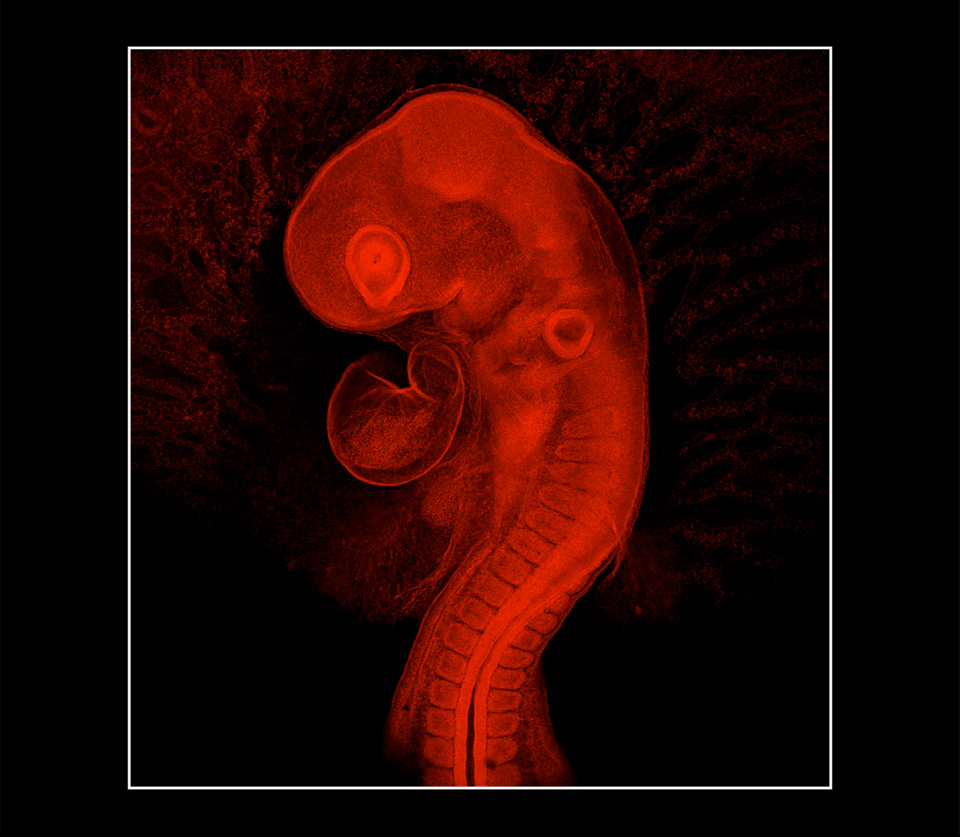
Chick Embryo at 48 Hours, 2018
Confocal photomicrograph
Excitation energy 520–580 nanometers
Rochester Institute of Technology
Rochester, New York, United States
This photomicrograph features a chick embryo. The sample was prepared 48 hours after fertilization. The specimen was approximately 3 millimeters from top to bottom.
