
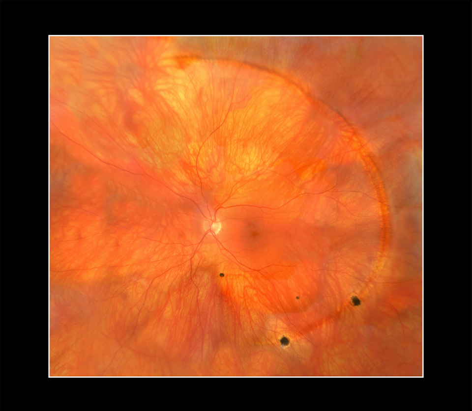
Joss Dimock
Worm Tracks in a Retina, 2018
Retinal fundus photograph
30 millimeters wide x 27 millimeters high; Topcon 50 EX fundus camera;
30 images taken at 50-degree views were stitched together using Adobe Photoshop;
overlapping edges were erased and some were distorted where blood vessels did not match up perfectly,
but the actual worm track was not distorted in any way;
composite image received a small amount of sharpening
Melbourne Health
Melbourne, Victoria, Australia
This is a composite retinal fundus photograph of a patient with worm tracks showing peripheral chorioretinal changes with pigment dispersion and focal areas of atrophy. The image was taken for clinical reasons.